
Florin Anghel
University Emergency Hospital Bucharest,
“Carol Davila” University of Medicine
and Pharmacy; Resident Physician
in Cardiovascular Surgery, PhD Candidate
The Technological Prelude in Medicine
Three-dimensional (3D) printing represents a cutting-edge technological solution through which two-dimensional (2D) image sequences—previously acquired using conventional imaging techniques such as ultrasound, computed tomography (CT), or magnetic resonance imaging (MRI)— are reconstructed with remarkable spatial precision. Following this virtual three-dimensional reconstruction, the imaging data are transformed into a physical object, materializing as a solid three-dimensional model. The utility of these physical models in cardiovascular surgery is exceptionally broad. Their applications range from the t9actile representation of highly complex anatomical structures such as cardiac tumors or patient-specific anatomical models routinely used in preoperative planning) to educational models designed specifically for medical simulation procedures.
The undeniable advantages of 3D-printing technology include a deeper understanding of the three-dimensional anatomy of complex structures, enhanced patient communication and informed consent processes, and the provision of a robust platform for procedural simulation and surgical planning.
Evolution and Current Status
Looking back over the past two decades, it is evident that 3D printing has achieved remarkable success within the medical field. A comprehensive analysis reported by Meyer-Szary et al. in 2022 examined scientific publications indexed in the PubMed database containing the keywords “3D printing” or “additive manufacturing.” The study highlighted an exponential growth in academic interest, with the number of publications increasing from just seven articles in 2000 to an impressive 4,817 scientific papers by 2020.
 Within cardiovascular surgery, this technology was initially employed primarily for the visualization of highly complex anatomical structures and for guiding reconstructive surgical procedures in patients with congenital heart disease. Today, in the era of endovascular therapies and increasingly complex clinical cases, 3D printing is extensively used for the pre-procedural planning of advanced endovascular interventions. Three-dimensional virtual segmentation of structures of medical interest significantly extends the diagnostic accuracy beyond the capabilities of traditional two-dimensional imaging. Although still considered a relatively recent technology, its applications in cardiac surgery are rapidly expanding and increasingly diverse.
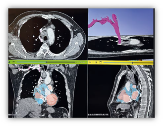
A representative example is the contrast-enhanced segmentation of the superior vena cava, highlighted in pink. The image demonstrates significant compression of the vessel lumen, strongly suggesting the presence of a cardiac tumor located in the right atrium.
Archives of the University Emergency Hospital Bucharest – Department of Cardiovascular Surgery, in collaboration with the 3D Printing Laboratory, Innovation and e-Health Center, Carol Davila University of Medicine and Pharmacy.
Preoperative Evaluation of Complex Anatomical Structures
In adult cardiac surgery, the principal applications of 3D printing during the critical preoperative planning phase include: Complex cardiac tumors, Ventricular aneurysms, Hypertrophic cardiomyopathy surgery and various coronary artery anomalies
A 3D model provides essential support by enabling surgeons to thoroughly understand the spatial relationships between a lesion and the surrounding anatomical structures, this insight is crucial for determining the most appropriate surgical approach.
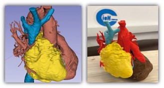
The image depicts a segmented model and a corresponding three-dimensional reconstruction of a giant right atrial angiosarcoma. The physical model allows precise visualization of tumor invasion into critical structures, including: the superior vena cava, the right atrium, the base of the right ventricle, the right coronary artery Source: Archives of the University Emergency Hospital Bucharest – Department of Cardiovascular Surgery and the 3D Printing Laboratory, Innovation and e-Health Center, Carol Davila University of Medicine and Pharmacy.
Patients who have experienced a myocardial infarction may develop a left ventricular aneurysm as a severe complication. In selected cases, this condition requires highly sophisticated surgical reconstruction. Within the surgical treatment algorithm for left ventricular aneurysms, a patient-specific 3D model provides substantial assistance in predicting the postoperative left ventricular end- diastolic volume. Such estimation is essential to avoid the development of a restrictive ventricular pattern or distorted ventricular geometry, both of which may subsequently result in severe mitral regurgitation. In this context, 3D printing serves as a direct pathway toward highly personalized and spatially guided surgical therapy.
The Role of 3D Models in Complex Endovascular Procedures
As previously emphasized, contemporary medicine is firmly rooted in the era of endovascular treatments. Among the most frequently performed procedures is Transcatheter Aortic Valve Implantation (TAVI).
3D printing has quickly proven its value in this field through the creation of tangible models that accurately replicate pathological structures within the area of surgical interest. These models fulfill several essential roles, such as:
- Enhancing communication between physician and patient
- Serving as invaluable educational tools for medical students and residents
- The biggest advantage is that it is enabling operators to perform pre-procedural simulations, including the deployment of prosthetic valves, in the initial phase, within the printed anatomical replica
Furthermore, virtual models provide highly accurate assessments of the so-called „ideal landing zone” for device placement. These sophisticated digital reconstructions can be seamlessly superimposed onto conventional fluoroscopic images.
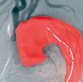
This image illustrates the superimposition of a virtual aortic endograft segmentation onto a fluoroscopic image, demonstrating the value of combining advanced imaging and 3D modeling technologies. Source: Archives of the University Emergency Hospital Bucharest – Department of Cardiovascular Surgery and the 3D Printing Laboratory, Innovation and e-Health Center, Carol Davila University of Medicine and Pharmacy.
Surgical Simulation and Educational Perspectives
Depending on the production technology and materials used, three-dimensional models can reproduce the physical properties of anatomical structures with remarkable accuracy. For example, soft silicone models can replicate not only the anatomy but also the biomechanical characteristics of structures such as the aortic root or mitral valve with exceptional fidelity. Their major advantage lies in allowing surgeons to both visualize and physically interact with patient-specific anatomy long before entering the operating room. Equally important, these models enable specialists to test various reconstructive strategies within a safe pre-procedural environment, without exposing patients to any risk.
Nevertheless, several obstacles continue to limit the widespread adoption of these innovative technologies. The primary challenges include the financial costs associated with production and the need for further refinement of currently available materials. Ongoing technological development is necessary to achieve mechanical and elastic properties that more closely resemble those of living human tissues.
In medical education, 3D printing offers a unique opportunity for the accurate reproduction of pathological conditions. The technology also facilitates the creation of fully customized simulators capable of reproducing a wide range of diseases across multiple medical specialties, generating surgical scenarios that closely mirror real-life operative situations.
Final Reflections
In conclusion, it is imperative to recognize that 3D-printing technology represents a genuine paradigm shift in the evolution of modern medicine.
This technological innovation is guiding the medical profession toward the adoption of fully personalized surgical interventions, with the ultimate goal of significantly improving therapeutic outcomes while ensuring the highest standards of patient safety.
